BIO 201 Human Anatomy & Physiology | ||||
| ||||
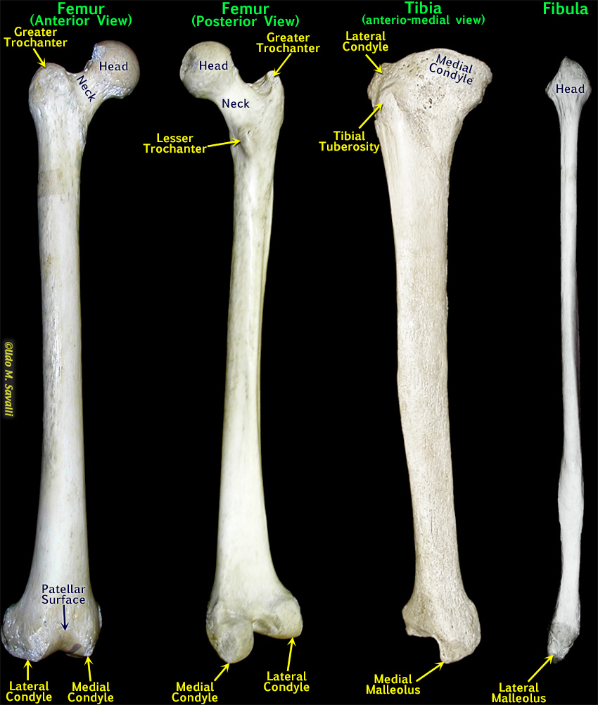
The markings of the femur, tibia, and fibulaIdentification: The femur is easily identified by its large size and distinct rounded head, allowing for a great range of movement. The femur can be separated from the humerus by the distinct neck separating the head from the rest of the bone. The tibia is also a large, heavy bone and thus potentially confused with the femur or humerus. Note that its superior end is rather flat-topped and lacks any sort of a rounded head. The fibula is distinguished by being longer and proporionately more slender than any other bone. Also note that it is enlarged and pointed at both ends. Determining side: For the femur, first orient it so that the rounded head is superior (up) and pointing medially (toward the body's midline). Then you will need to determine the anterior vs. posterior side. Look for the patellar surface, which is anterior. Also note how the articulating surfaces of the condyles extends far back on the posterior side (since the knee bends back but not forward). For the tibia, first orient it so that the larger, flatter end is superior (up). The tibial tuberosity and shin (a ridge) are on the anterior (front) of the bone. Finally, the medial side can be determined by the medial malleolus (remember that the malleoli bracket the ankle and since the tibia is the medial bone of the lower limb, its malleolus must be medial). A right tibia is shown. You do not need to distinguish left and right on the fibula. |
|
This page last updated 18 August 2019 by Udo M. Savalli () Images and text © Udo M. Savalli. All rights reserved. |