BIO 370 Vertebrate Zoology | ||||
Vertebrate Anatomy | ||||
Labeled Tissues
| |||||||||||||||||
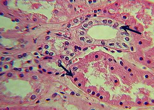
Simple cuboidal epithelium (from kidney). Arrows indicate nuclei. |
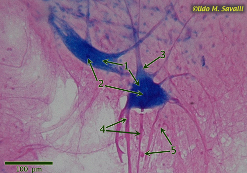
Motor neurons. 1 = cell body; 2 = nucleus; 3 = axon; 4 = dendrites; 5 = glial cell nuclei |
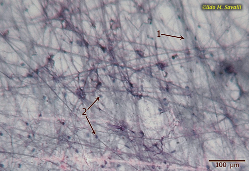
Loose (or areolar) connective tissue. 1 = collagen fiber; 2 = nuclei of fibroblasts |

Fibrous (or dense) connective tissue (from tendon). 1 = fibroblast |
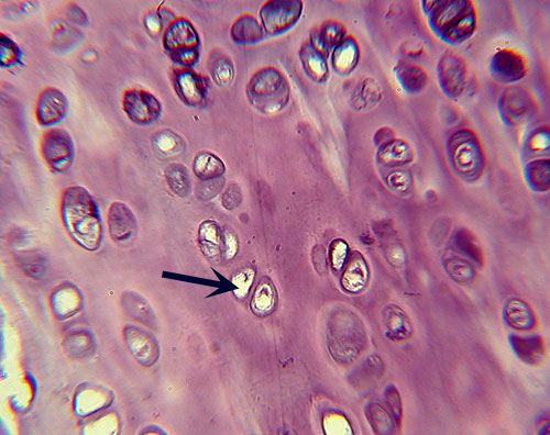
Hyaline cartilage. Arrow indicates chondrocyte within lacuna |
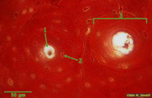
Compact bone, showing: 3 = osteon (Haverian system); 1 = Haversian canal; 2 = osteocyte in lacuna |
|
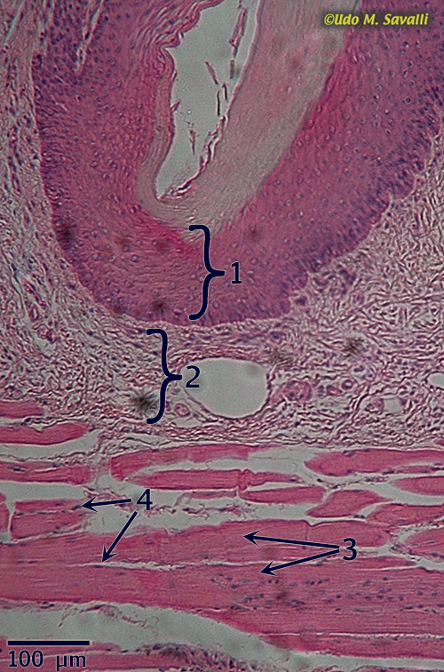
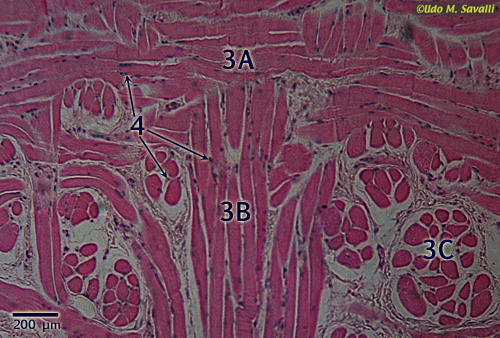
Left and below: section through mammal tongue showing:
|
| |
|
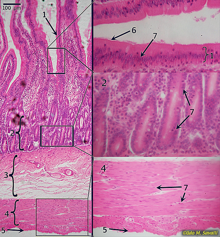
Section through small intestine showing:
|
|
This page last updated 8 September 2010 by Udo M. Savalli () Images and text © Udo M. Savalli. All rights reserved. |