|
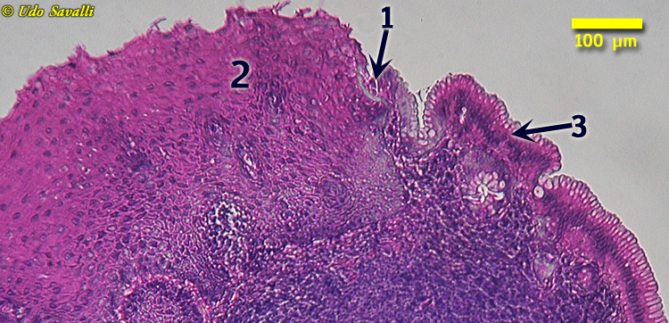
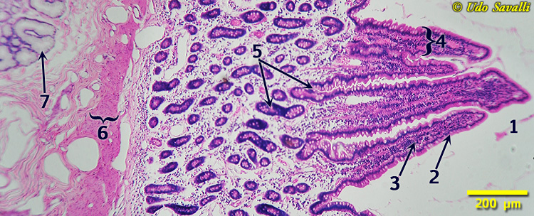
Gastroesophageal Junction
- Gastroesophageal Junction
- Stratified squamous epithelium of esophagus
- Simple columnar epithelium of stomach
|
|
|
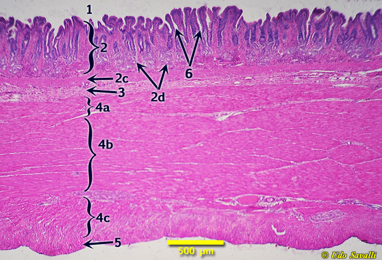
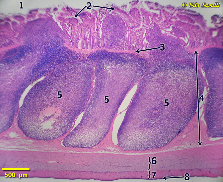
Stomach
- Lumen
- Mucosa
- Simple columnar epithelium
- Lamina propria (areolar CT)
- Muscularis mucosae
- Gastric glands
- Chief cells
- Parietal cells
- Submucosa
- Muscularis externa
- Oblique layer
- Circular layer
- Longitudinal layer
- Serosa
- Gastric pits
|
|
|
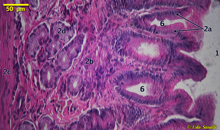
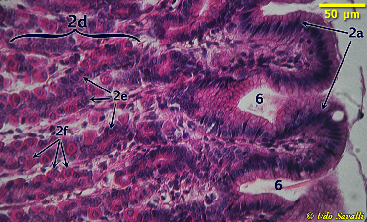
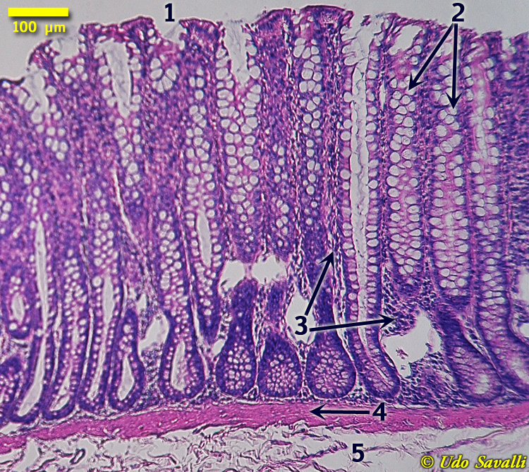
Duodenum
- Lumen
- Simple columnar epithelium
- Lamina propria
- Villus
- Intestinal crypt
- Muscularis mucosae
- Duodenal gland
|
|
|
Ileum
- Lumen
- Villi
- Muscularis mucosae
- Submucosa
- Aggregated lymphoid nodules (Peyer's patches)
- Muscularis externa: circular layer
- Muscularis externa: longitudinal layer
- Serosa
|
|
|
Colon (Large Intestine)
- Lumen
- Goblet cells (of columnar epithelium)
- Lamina propria
- Muscularis mucosae
- Submucosa
|
|